In 2014, at the age of 19, Leonardo Lima had just dropped his mother off at work in the city of Macaíba (RN), when he was approached by robbers. As he accelerated his motorcycle in an attempt to escape the robbery, he was shot several times by the robbers, resulting in a complete spinal cord injury. Complete spinal cord injury is diagnosed when there is no voluntary movement below the level of the trauma. Part of the central nervous system, the spinal cord is responsible for conducting signals related to the brain's sensitivity to the rest of the body. Until the 1990s, it was believed that, in cases like Leonardo's, when suffering a severe trauma, communication between the spinal cord and the brain would be completely interrupted. Studies on cadavers, however, have revealed that this is not the case.
In 1998, Byron Kakulas, a physician and one of the founding directors of the Neuroscience Institute at the University of Western Australia, presented data that revealed the preservation of up to 27% of the so-called “white area” of the spinal cord, where the axons, the part of the neuron responsible for propagating signals to the brain and back to the spinal cord, are concentrated. This means that, contrary to what was previously thought, communication between the area below the lesion and the brain was not completely interrupted.
With the advancement of technology, the perception that a complete spinal cord injury might not be so complete gained ground in the scientific world, especially with the consolidation of studies on neuroplasticity – the ability of our neurons to adapt both anatomically and functionally to certain situations.
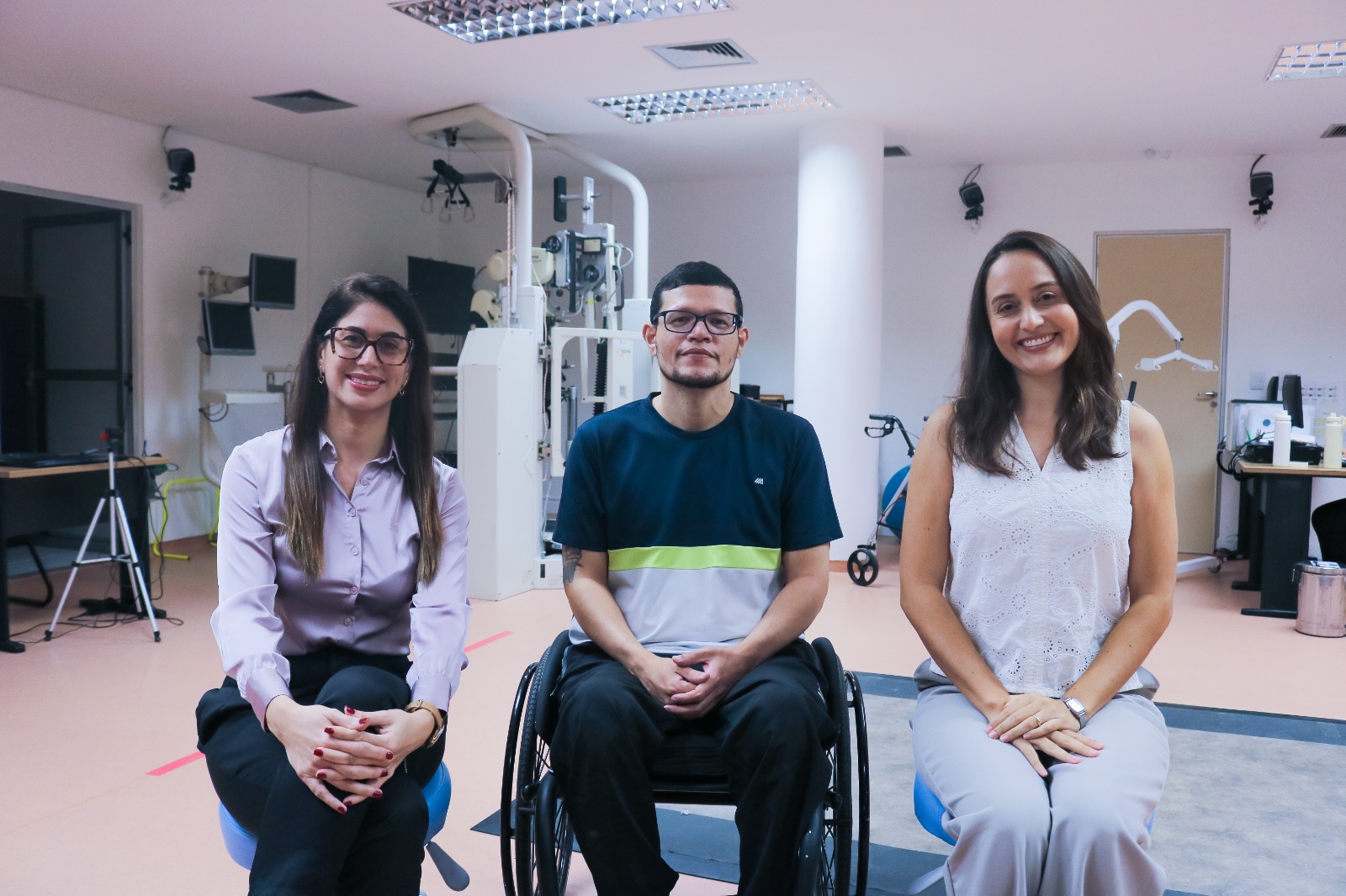
Camila Simão, a member of the physical rehabilitation clinic at the Santos Dumont Institute (ISD), joined those who sought to study how these remaining connections could influence the rehabilitation process – and what tools could be used to visualize the existence of these connections. During her PhD research at the Federal University of Rio Grande do Norte (UFRN), Camila investigated the use of electromyography as a tool to identify the preservation of these remaining pathways in patients diagnosed with complete and chronic spinal cord injury.
The research was also published in the form of an article in the European Journal of Physical Rehabilitation Medicine, in which the physiotherapist presents the study carried out with the patient Leonardo Lima, in which the electromyography technique was able to identify remaining connections in the spinal cord.
Electromyography
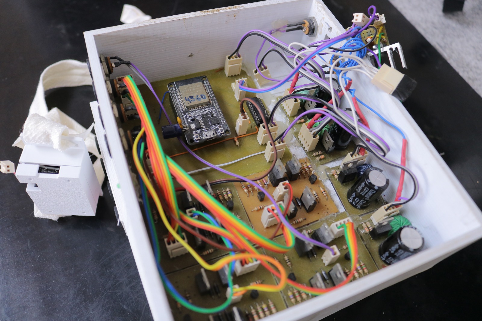
Electromyography is a type of neurophysiological examination in which small sensors (surface electrodes) are placed on the muscles to measure the electrical activity of muscle contraction. This is possible because the contractions and movements we make in our body generate this type of current, which can be measured by the equipment. The technique was first used in the 1970s and, since then, its uses have evolved and the examination has come to have a series of applications.
Unlike other techniques that can be used to measure muscle activity, it is non-invasive and much cheaper than other equipment. That is why Camila Simão decided to use the technique to try to bring it to rehabilitation clinics. “There are already other effective ways to perform this measurement, but they are expensive, complex or cause discomfort for patients. With electromyography, we were able to perform an analysis at a much lower cost, so that this practice can be adopted not only for scientific research, but in the clinic itself,” explains Simão.
This is how Leonardo Lima was able, for the first time since he suffered a spinal cord injury, to visualize the activity of his muscles. With the surface electrodes installed, he was placed standing on a platform that supported him. Beside him, Camila Simão gave commands for him to imagine the movement as strongly as possible. Off-screen, the force applied by Leonardo seemed to have no effect on the movement of his legs. On the computer that recorded the movements, however, the reality was different: with each thought of contraction, it was possible to see an effect on the monitor, where the signal increased with the effort of the movement.
For the researchers, the findings point to, as Camila said, “a new paradigm in the process of evaluating and rehabilitating this population” given the possibility of neuroplasticity. However, this was not the only practical effect identified by the research. Leonardo’s visualization of the movements through electromyography, for example, gave him even more desire to dedicate himself to rehabilitation. “When I saw that sign going up, I wanted to try more, more and more. It’s another type of visualization, another type of understanding that we start to have of our body and the life within it,” he declared.
Text: Mariana Ceci / Ascom – ISD
Photograph: Mariana Ceci / Ascom – ISD
Communication Office
comunicacao@isd.org.br
(84) 99416-1880
Santos Dumont Institute (ISD)
It is a Social Organization linked to the Ministry of Education (MEC) and includes the Edmond and Lily Safra International Institute of Neurosciences and the Anita Garibaldi Health Education and Research Center, both in Macaíba. ISD's mission is to promote education for life, forming citizens through integrated teaching, research and extension actions, in addition to contributing to a fairer and more humane transformation of Brazilian social reality.